
Accurate protein quantification is a cornerstone of molecular biology and biochemistry protocols. Selecting the optimal assay depends on your sample composition, protein concentration, presence of detergents or interfering agents, and required sensitivity.
Here is a comprehensive comparative guide to four classic protein quantification methods: UV Absorbance (A280), Bradford Assay, Lowry Assay, and BCA Assay.
Part 01: UV Absorbance Method (A280)
Principle:
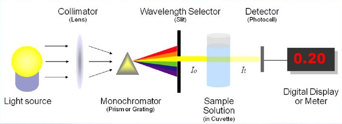
Aromatic amino acid residues—primarily Tryptophan (Trp), Tyrosine (Tyr), and Phenylalanine (Phe)—absorb UV light at 280 nm. Based on the Beer-Lambert Law ($A = \varepsilon \cdot c \cdot l$), absorbance at 280 nm is directly proportional to protein concentration ($c$). On average, an absorbance ($A_{280}$) of 1.0 corresponds to approximately 1.0 mg/mL of protein.
Advantages:
Rapid & Easy: Direct measurement using a spectrophotometer without incubation or chemical reactions.
Non-Destructive: The sample remains chemically unaltered and can be fully recovered for downstream assays.
No Reagents Needed: Eliminates the cost and preparation of chemical dyes or color reagents.
High Dynamic Range: Suitable for concentrated protein samples.
Limitations:
Nucleic Acid Interference: DNA and RNA absorb strongly at 260 nm and moderately at 280 nm, distorting readings if samples are contaminated.
Composition Dependency: Proteins with unusually high or low Trp/Tyr content deviate significantly from the average extinction coefficient ($\varepsilon$).
Low Sensitivity: Inaccurate for ultra-low concentration samples.
Part 02: Coomassie Brilliant Blue Method (Bradford Assay)
Principle:
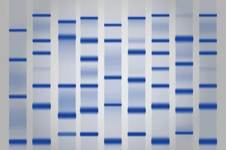
Coomassie Brilliant Blue G-250 dye binds to protein molecules under acidic conditions. Upon binding (primarily via hydrophobic and ionic interactions with basic amino acids like Arginine and Lysine), the dye shifts from a red/brown form ($A_{max} = 465\text{ nm}$) to a blue form ($A_{max} = 595\text{ nm}$). Absorbance at 595 nm correlates directly with protein concentration.
Advantages:
High Sensitivity: Detects concentrations as low as 1–10 µg/mL.
Fast Workflow: Single-reagent addition with rapid color development within 2–5 minutes.
Chemical Compatibility: Highly resistant to standard salts, buffers, and reducing agents (e.g., DTT, Mercaptoethanol).
Limitations:
Detergent Incompatibility: Strongly interfered with by detergents like SDS or Triton X-100.
Protein-to-Protein Variation: Varies based on basic amino acid content; requires a standard curve (typically Bovine Serum Albumin, BSA).
Cuvette Staining: Dye binds to plastic and glass cuvettes; disposable cuvettes or microplates are recommended.
Part 03: Lowry Protein Assay
Principle:
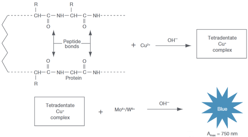
A two-step reaction combining the biuret reaction with the reduction of Folin-Ciocalteu reagent. Copper ions ($Cu^{2+}$) bind to peptide bonds under alkaline conditions and reduce to $Cu^+$. Reduced cuprous ions then react with Folin-Ciocalteu reagent (phosphomolybdic-phosphotungstic acid), producing a intense blue complex measured at 750 nm (or 660 nm).
Advantages:
High Sensitivity & Precision: Highly reproducible with lower detection limits.
Less Composition-Dependent: Reacts primarily with the peptide backbone rather than specific amino acid side chains.
Limitations:
Complex Protocol: Involves multi-step reagent additions and precise incubation timing.
Interference-Prone: Highly sensitive to EDTA, Tris buffers, reducing agents, and non-ionic detergents.
Reagent Instability: Folin-Ciocalteu reagent must be freshly prepared and protected from light.
Part 04: Bicinchoninic Acid Assay (BCA Method)
Principle:
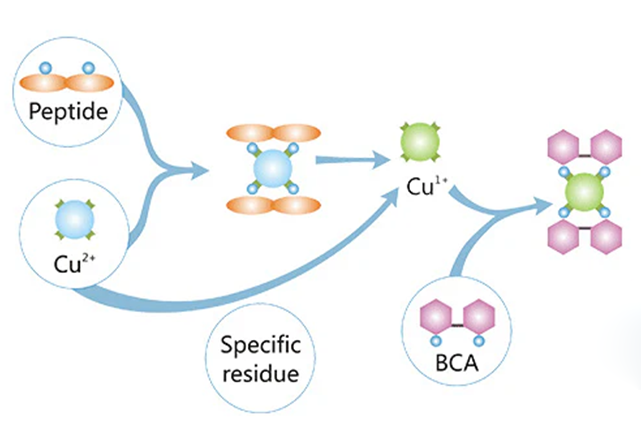
Combines the reduction of $Cu^{2+}$ to $Cu^+$ by peptide bonds under alkaline conditions with the chelation of $Cu^+$ by Bicinchoninic Acid (BCA). Two molecules of BCA react with one $Cu^+$ ion to form a purple-colored complex with strong absorbance at 562 nm.
Advantages:
High Detergent Tolerance: Fully compatible with up to 5% SDS, Triton X-100, and NP-40, making it ideal for cell lysate samples.
Broad Linear Range & Stability: Excellent linearity with a stable color complex that allows flexible measurement windows.
Ease of Use: Stable, ready-to-use liquid reagents; simpler than the Lowry method.
Limitations:
Incompatible with Reducing Agents: DTT, DTT-like compounds, or DTT/2-ME destroy the $Cu^{2+} \rightarrow Cu^+$ reaction.
Temperature Dependent: Requires incubation at 37 °C or 60 °C to accelerate color development.
Method Selection Summary Matrix
| Assay Method | Key Mechanism | Optimal Wavelength | Detergent Tolerance | Reducing Agent Tolerance | Typical Sensitivity |
|---|---|---|---|---|---|
| UV A280 | Aromatic Residues (Trp/Tyr) | 280 nm | High | High | High Concentration (0.1–3 mg/mL) |
| Bradford | Coomassie G-250 Binding | 595 nm | Poor | Good | High (1–20 µg/mL) |
| Lowry | Biuret + Folin Reduction | 750 / 660 nm | Moderate | Poor | Very High (1–100 µg/mL) |
| BCA Assay | $Cu^+$ Chelation by BCA | 562 nm | Excellent | Poor | High (0.5–20 µg/mL) |
About A-Gen
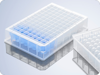
Founded in 2009, A-Gen Biotech is dedicated to the R&D and manufacturing of high-quality laboratory consumables for life sciences, biotechnology, and clinical diagnostics. Operating under strict ISO 13485 and CE quality standards, A-Gen provides trusted, high-performance solutions—including microplates, spectrophotometer cuvettes, and micro-volume tubes—to researchers worldwide.
Choose A-Gen, Choose Quality.